Embolization of uterine fibroids
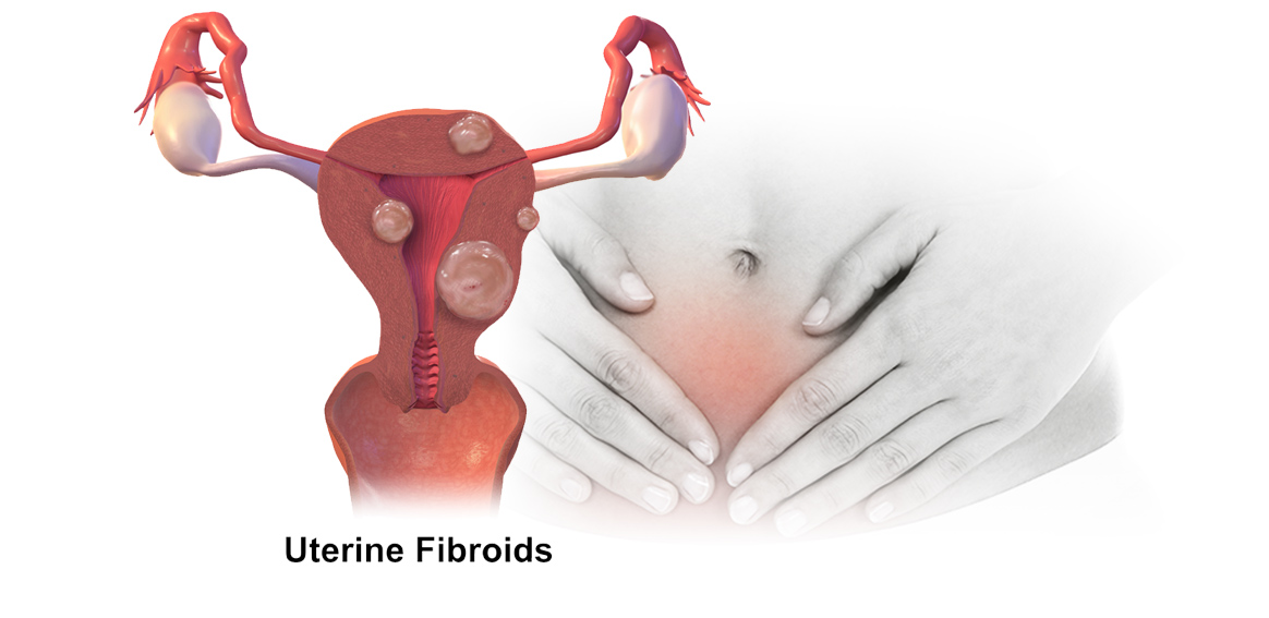
Uterine fibroids
Fibroid is the muscle tissue of uterine that grows abnormally in the form of a lesion. The lesions can grow in different places, but they grow most commonly in the uterine wall. The size of these tumors can be more than 10cm and they may be numerous.
Fibroids can be seen in more than 30% of women an can develop without any symptoms or discomfort. It can be observed randomly on ultrasound and disappear on its own after menopause.
Follow-up and evaluation of uterine fibroids
In some women, uterine fibroid leads to pelvic pain, irregular menstrual bleeding, and urinary disorders. It rarely leads to infertility and, in very rare cases, uterine fibroids become malignant and cancerous.
The most common reason due to which patients with uterine fibroids are admitted is irregular bleeding, which sometimes leads to severe anemia.
New non-surgical treatment of uterine fibroids (uterine fibroid artery embolization)
Uterine fibroid artery embolization is one of the methods for treating uterine fibroids, which has been of great interest recently.
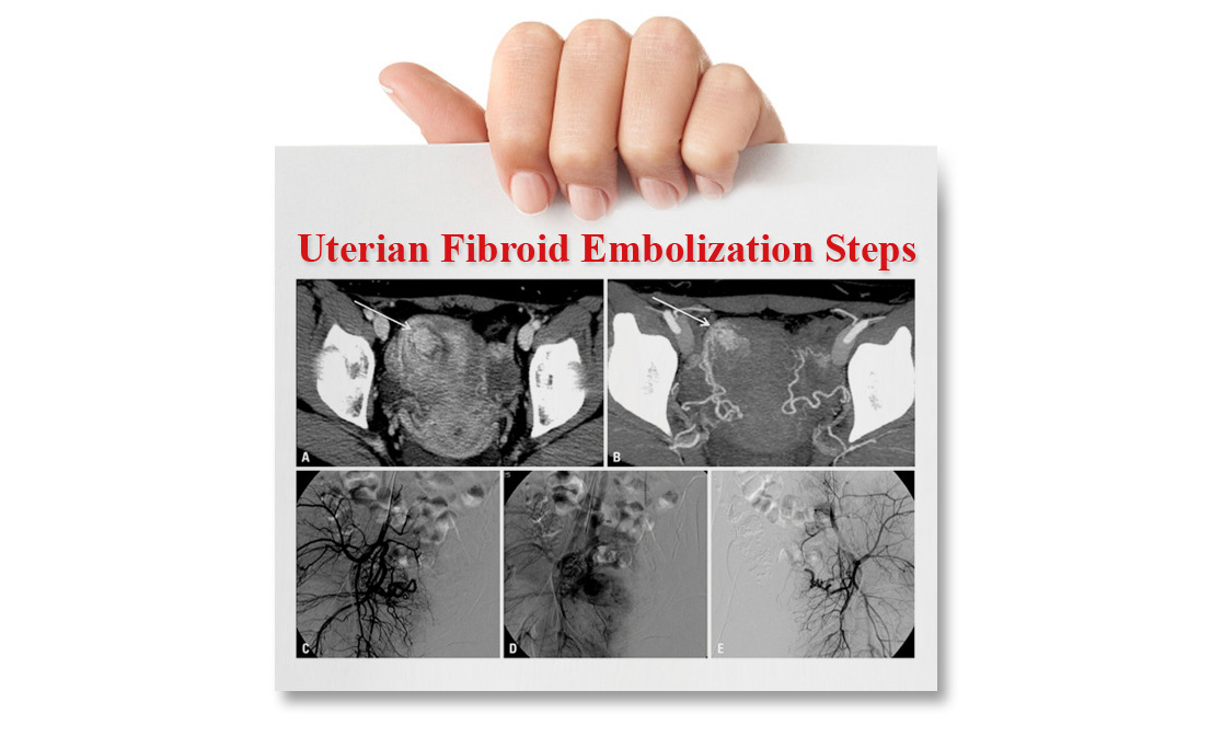
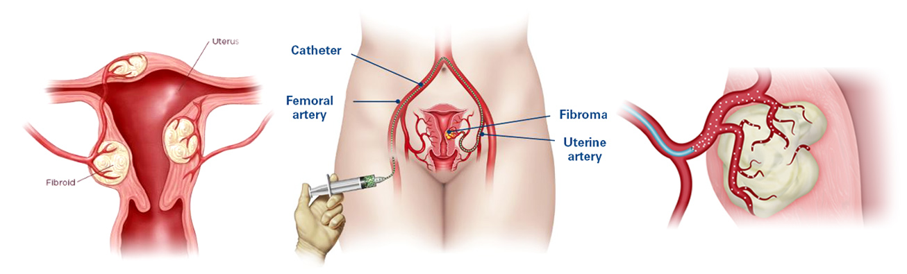
In this method, a combination of several blockers will be injected into the uterine fibroid artery, leading to the blockage of the blood supplying vessels. Thus, with blood and oxygen supply cut off, uterine fibroids tumor will be necrosis and disable. About 90 percent of people who have been treated observed a significant improvement in their symptoms. Radiologists recommend MRI for determining whether or not the fibroid is a candidate for embolization.
MRI helps to diagnose the vessels feeding the uterus fibroid, thereby enabling better treatment.
Embolization is recommended in the following cases:
– Fibroids that cause severe bleeding
– Fibroids causing pain and pressure in the bladder and rectum
– Unwillingness to hysterectomy
– Unwillingness to pregnancy
Required preparations for uterine fibroid artery embolization
– Fasting for 6 hours
– Having the previous medial records
– Having coagulation blood test results
– Having a companion for doing the administrative affairs
How uterine fibroid embolization is performed
After verification of the medical records by the radiologist under the guidance of imaging devices and local anesthesia, a catheter will be inserted into the uterine fibroid artery through the right groin. Then, a combination of several blockers will be injected into the intended artery and the imaging process will always be checked by the device.
Finally, the catheter will be removed from the artery and bandage will be applied to the incision site. Then, bleeding will be prevented by placing an Angio-Seal and sandbag.
Uterine fibroid artery embolization will be performed without incision and only with a cut of few millimeters the trace of which will disappear after a short time.
Advantage of uterine fibroid artery embolization
This procedure is performed without anesthesia and has much less interference than surgery-
The patient will return to normal life as soon as possible-
Bleeding in this method is much lower than surgery-
Recurrence percentage is very low-