


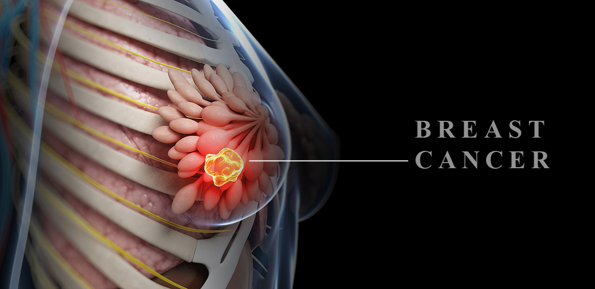
Breast Cancer
Breast cancer is one of the most common malignant cancers in women that can be diagnosed at an early stage through some periodical tests.
Sings and Symptoms of breast cancer
– Lumps in the breast
– Nipple sensitivity
– Thickening of the breast or armpit skin
– Skin irritation of changes, such as dimpling, puckering, and dimpling
– Warmth, redness, and swelling associated with acne and breast skin rash
– Discharges of physical changes (such as nipple turning inward, persistent pain or changes in the size and shape of the breast)
People who should be more careful include:
– Women who have a family history of breast cancer
– Women with early menstruation and late menopause
– Women who have experienced their first pregnancy at above 30
– Smoking, obesity after menopause, and high fat consumption are among other factors
Mammography
Mammography is a digital X-ray image of the breast for early diagnosis of non-palpable cancers. Mammography can detect slow-growing malignant breast tumors at least 2 years before reaching the palpable size.
The proper age for mammography
The best age for mammography is over 35. At older ages, mammography is recommended annually or every two years according to the patients condition.
The necessity of mammography
mammography can detected small anomalies in breast tissue that the patient or the doctor cannot touch and determine during the examination. The first mammography, as the base image, will help the radiologist detect any changes through comparing it with the next mammography. One of the points the radiologist is looking for in mammography is “micro calcification” or fine particles of calcium deposits, which are show in the image as small white dots. In many countries, mammography is performed as a screening test on all women.

Digital mammography and its advantages to conventional mammography
Less amount of the used radiation compared to conventional mammography
Very high quality of images compared to conventional mammography
Zooming digital images as required in high quality and high precision
Recording and storing digital mammography images on CD
Preparing digital images in less than 10 seconds compared to conventional mammography

Breast Ultrasound
Using ultrasound, the breast characteristic can be examined and breast lumps detected.
Based on the nature of these lumps, benign lumps (such as cysts or fibroadenomas) or the lumps suspected with malignancy can be detected. Appropriate and accurate ultrasound requires advanced equipment as well as experienced doctors who are expert in this field.
Note:
For better diagnosis and more precise results, ultrasound and mammography will be performed in a complementary manner. That is because they both have a great contribution to diagnosis of the suspected case.
MRI-guided Biopsy
In this method, a sample of breast tissue will be biopsied under MRI guidance. The position of breast tissue lesion and location of the biopsy needle for taking sample will be determined under MRI guidance. Biopsy needle will be inserted into lesion per-cutaneously and the suspicious tissue sample with be biopsied. This technique is less invasive than surgery and has no X-ray compared with mammography-guided biopsy. On the other hand, it has a much higher accuracy in determining the malignant area for biopsy. Patient recovery time after biopsy is shorter, and the patient quickly returns to normal daily activities.
Ultrasound-guided Biopsy
Using this technique, pieces of tissue from the suspicious lump in the breast will be biopsied and examined for pathology under ultrasound guidance and under local anesthesia without any need for surgery.

MRI of the Breast (MR Mammography)
MR mammography is the most accurate, sensitive method for detecting breast tumors that can be performed through a collection of the following equipment:
– Dedicated breast coils
-Drawing wash-out and wash-in curves
– Evaluating the response to chemotherapy after 1 to 2 sessions of the treatment
– Dynamic injection and examination of tumor perfusion
– Breast diffusion images
Radiofrequency Ablation (RFA) of Breast Lumps
RFA ablation is a minimally invasive, non-surgical technique which has been recently of interest to the physicians for the treatment of breast lesions and some beast cancers. In this procedure, breast lesions and tumors will be destroyed and ablated using radio waves in a minimally invasive manner.
RF Neddles
(Electrode) will be inserted into the breast lesions percutaneously and radio waves be transferred to the tumor through these electrodes and the tumor or lesion will be destroyed as a result of the produced heat.
