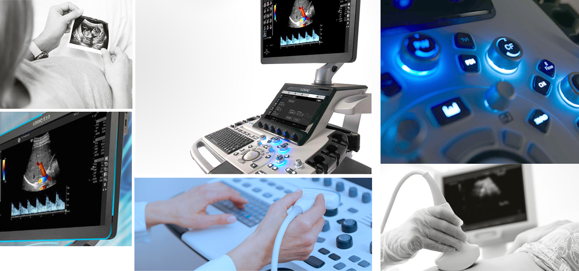


As we know, in X-ray imaging methods such as simple X-ray or CT scan, the body is exposed to a certain amount of ionizing radiation. However, X-ray are not used in ultrasound. The waves used in ultrasound are sonic waves, and they are in fact sound which is not harmful to the body. These waves, which are exactly like sound, but they are not audible by the human ear because of their high frequency. Nevertheless, they have properties of the sound; that is, they are rebounded after hitting obstacles.

Ultrasound imaging is not painful and the patient feels no discomfort during the procedure.
In ultrasound, most soft tissues of the organs can be seen and examined. Thus, ultrasound is not a suitable method for detecting bone problems. However, it can examine the problems in the muscles of ligaments and tendons and many other tissues.
Ultrasound my be two-dimensional or three-dimensional. Meanwhile, the ultrasound images of organs can be seen while in motion and activity.


There is a special type of ultrasound called Doppler ultrasound. This imaging technique works on the basis of the Doppler phenomenon and aims to check the amount and speed of blood flow in the body veins and arteries. This method is more used for assessing the possibility of clots in the deep veins of the leg.
FNA
It is the gold standard method for distinguishing thyroid benign lesion from malignant lesion that provides helpful distinguishing information of 80% of single nodules.
FNA has increased in recent years because of the high level of thyroid nodules diagnosed by ultrasound.
Hopefully, using thyroid elastography and Doppler examination unnecessary FNA on the study population will be reduced.
Elastography can be used in ultrasound for assessing tissue elasticity because it has the potential to detect cancer in single nodules of thyroid.