
This device provides suitable conditions for imaging even of children by ability to adjust different positions, change the contrast, brightness, and gamma-ray. Sensors used in this device are CT-type sensors that have a major and direct effect on increasing the quality of dental therapies.
In addition to the increased quality of imaging, another advantage of this device is the reduced time needed for general imaging of the teeth, The imaging duration has reduced from 17 seconds to 10 seconds compared to the old system of Panorex. Meanwhile, the Panorex image can be seen on the monitor immediately after imaging, and a better image can be created in case of need.
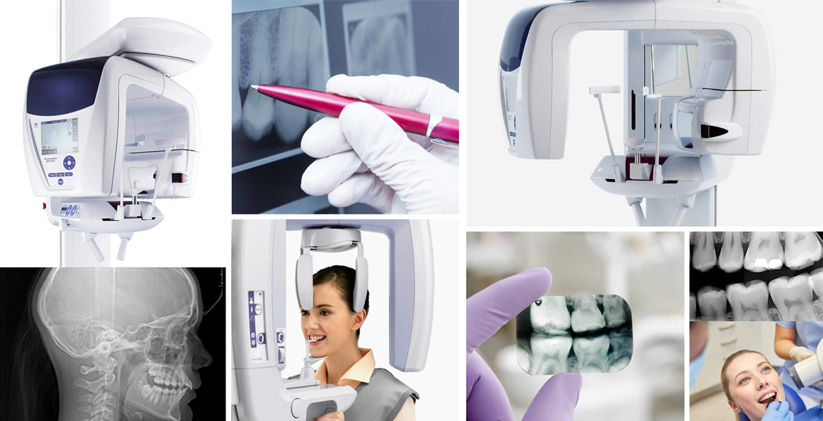
This feature has eliminated film waste.
Other specialized features of this device are as follows:
The image taken using the normal Panorex is usually used for seeing an overview of both jaws, the number of teeth and the roots in the jaw. However, in digital Panorex device, the resolution of a zoomed image is high enough to see the root length of all single teeth clearly, like periapical (single tooth) X-ray. That is periapical (single toot) X-ray of all teeth can be prepared through radiating X-ray only once.
Another special feature of this device is the accurate measurement of the decay of each tooth for restoration and root canal therapies.
Meanwhile, the length of root is also important in implant and orthodontic treatments and the use of this device enables online measurement and observation by the doctor.
Panorex Services :
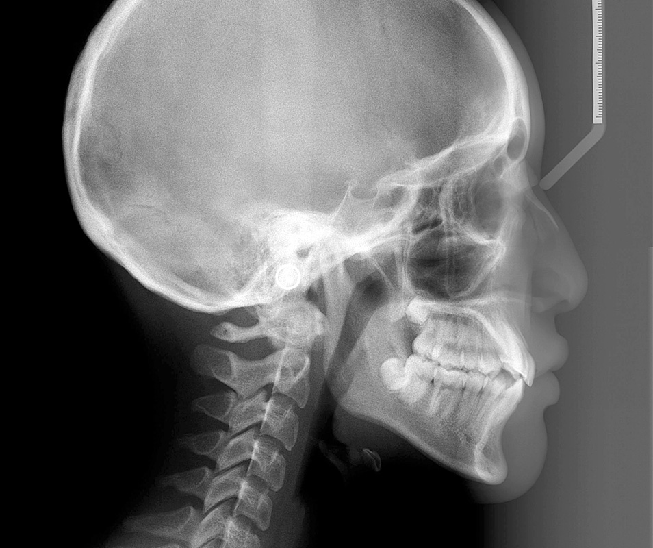
Panorex X-ray, cephalometry in two directions and TMG
Parallel and periapical bitewing in adults
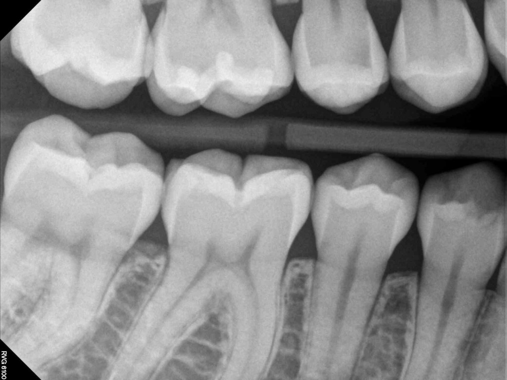
It is a type of X-ray machine that can provide highly precise images of one or more limited teeth to show the full status of the tooth and its surrounding tissue. It is called periapical because it well illustrates the lesion around the apex or root tip. However, this technique is also used for examining the bony crest edges around the teeth as well as he interdental decays.
However, the bitwing procedure is used in the latter case (interdental decay).
n addition, occlusal images of each jaw can also be obtained using larger films.
BITEWING :