

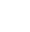
Interventional Radiology
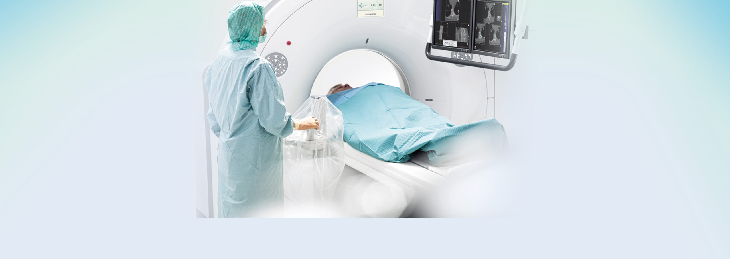
Interventional radiology, briefly referred to as IR, is a specialized field of radiology performed with the help of (CT, ultrasound, MRI, fluoroscopy) devices and special techniques and the doctor’s required expertise and skills, and diagnosis and treatment of the patient.
Many diseases that once required extensive surgery are now easily treated with the help of this technique and without surgery. Almost all interventional procedures can be performed by making a small incision on the skin and, as a result, the complications are much less.
The advantages of interventional measures:
Reduced bleeding during the surgery
Reduced infection rates
Reduced hospitalization period
Reduced risk of surgery
Reduced pain
Recovery in a short time, often without anesthesia
Interventional radiology is continuously progressing and in recent years, experts in this field have presented valuable services to the medical community through required innovations and inventions.
Nephrostomy
It is the insertion of a special catheter into the kidney system in order to divert the urinary tract in patients with ureteral obstruction, which is performed with the help of ultrasound and fluoroscopy under local anesthesia. The end of nephrostomy catheter is suture-fixed to the skin to avoid going off of the nephrostomy catheter.
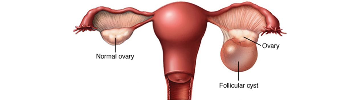
Drainage of various types of cysts
Abdominal, ovarian, breast, liver, etc. cysts will be drained using imaging devices under local anesthesia by special needles, and they are sent to laboratory for cytology.
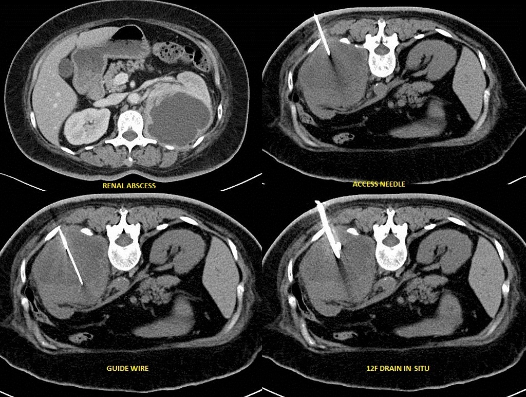
Drainage of abscess
It is the catheterization of the abdominal and pelvic abscess for draining the abscess and infection using the imaging device under local anesthesia. The catheter is connected to the bag by an interface and drainage is established. This catheter is fixed on the skin by sutures and fixator to hold the catcher and not to let it go off. After the full drainage of abscesses and imaging for full assurance, the catheter will be removed from the body.
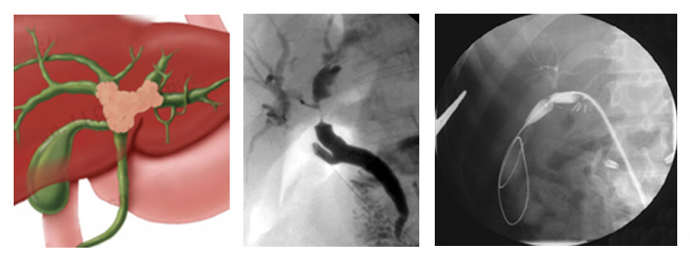
Bile drainage
For patients in whom bile duct obstruction causes jaundice and itching, a special catheter will be inserted into the obstruction duct using the imaging device under local anesthesia. In cases where severe obstruction blocks the way for passing a catheter through the length of stenosis, the end of the catheter will be connected to a bag and bile will be driven out of the abdomen to prevent from the aggregation of bile in the biliary ducts.
Biliary duct stenting
Metal stenting is another treatment for removing biliary obstruction, which is performed using the imaging device under local anesthesia. That is, the stenosis location is first detected by the imaging device. Then, the obstructed path will be unblocked by special spring and balloons and the metal stent will be put in stenosis so that the natural course of bile into the intestine would open.
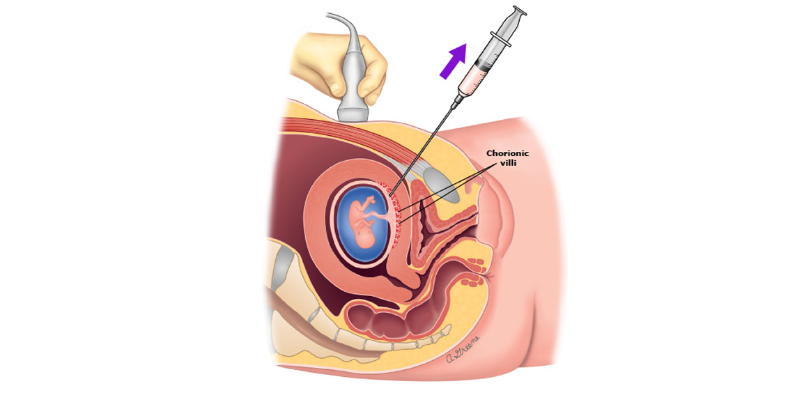
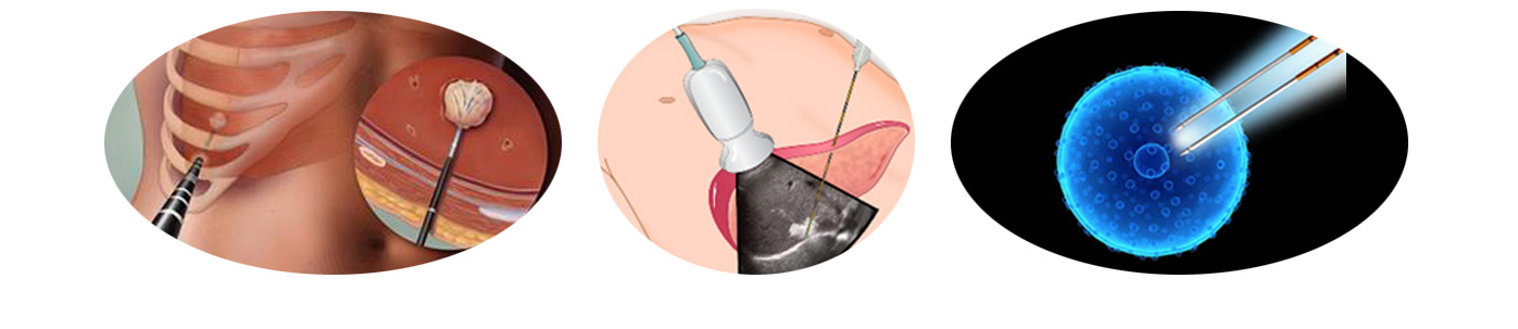
Sampling from the placenta
CVS or sampling from placenta villus is performed for detecting prenatal genetic abnormalities and is recommended to be done between weeks 11 and 14 of pregnancy. This sampling is performed using an ultrasound device under local anesthesia.
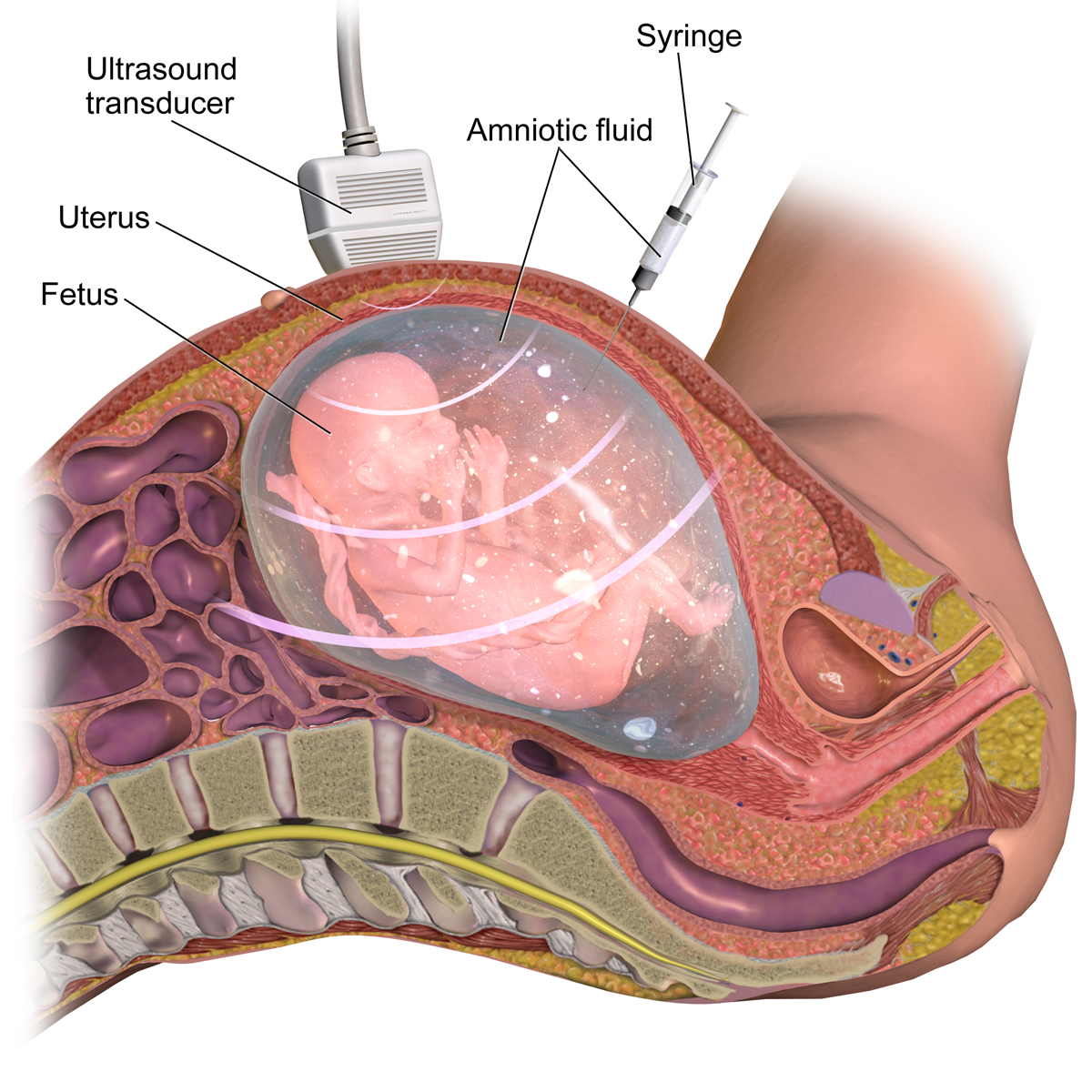
Amniocentesis
Amniocentesis is the aspiration of the fluid around the fetus for prenatal diagnosis, and is performed using an ultrasound device. It is recommended to be done between weeks 14 and 18 of pregnancy.
Ascites drainage
Some cancer patients may suffer from the accumulation of free fluid in the abdomen or pelvis, in which case ascites must be drained by inserting a special catheter into the desired area because it causes asthma and constipation. Ascites drainage is performed using imaging devices and under local anesthesia and the end of catheter will be fixed to the skin surface by sutures and fixators so that it would not go off. After the full drainage of ascites, the catheter will be removed from the body.

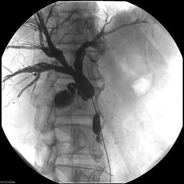
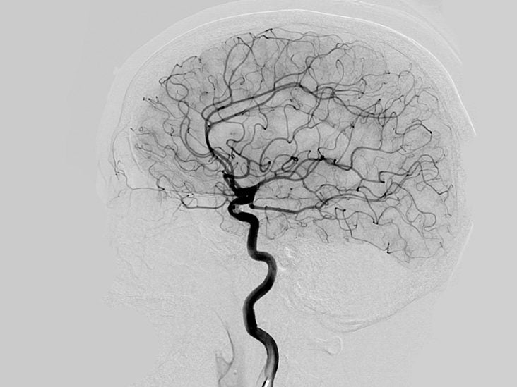
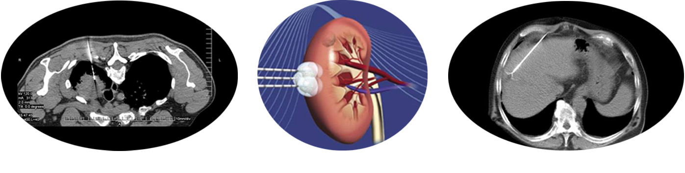
Angiography
Angiography is another new imaging method for diagnosing vascular abnormalities (obstruction, aneurysms, EVM, etc.). Angiography is performed using imaging devices under local anesthesia.
That is, a special catheter is inserted into the desired arteries through the right groin and the required diagnostic images will be taken by contrast agent injection.
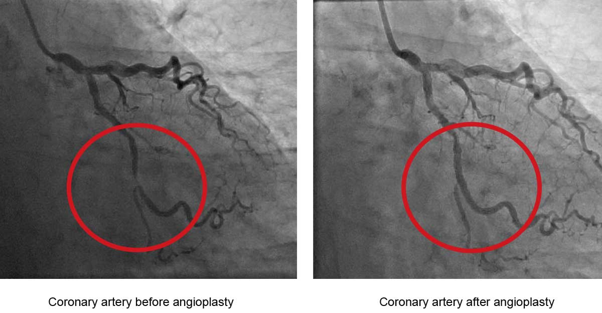
Angioplasty
In this procedure, a special balloon catheter will be used for removing the obstruction and narrowing of the arteries. That is, using imaging devices and under local anesthesia, a special catheter is inserted into the desired arteries through the right groin and the required diagnostic images will be taken. Then, the angiography balloon will be inserted into the artery stenosis and stenosis will be unblocked with air injection into the balloon.
Coiling damaged arteries
In cases where there is a vascular lump in the artery path (which is usually congenital and grows very slowly), it impairs the quality of life. In these circumstances, it is necessary to take certain remedial measures. That is, first, using imaging devices, a special catheter will be inserted into the vascular lump through the right groin. After angiography and obtaining the required images, the lump vessels will be ablated by specials springs and adhesives and a rupture that can lead to bleeding will be avoided.

Lump ablation of liver, lung, kidney, etc. through radio frequency technique
Radio frequency ablation
Radio frequency ablation technique ablates the tumors of the liver, lung, bone, breast, etc. at high temperatures, destroying cancer cells in the area. This will be performed using imaging devices and under anesthesia and is considered an outpatient procedure due to its short-term hospitalization.
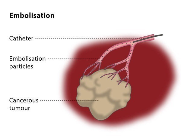
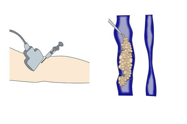
Embolization
It is a method for blocking blood vessels that supply blood to the intended points such as uterine fibroids, hemangiomas, and vascular lumps that cause a lack of blood supply to the desired area and, thus, deactivate the desired area. That is, first, using imaging devices, a special catheter will be inserted into the desired artery through the right groin. After angiography and obtaining the required images, a micro-catheter will be inserted into the artery. Then, artery embolization will be performed via injection of blocking substances.
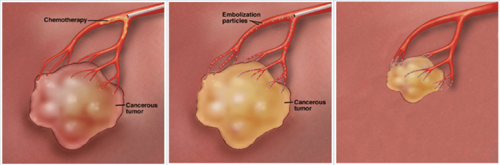
Cemoembolization
It is a method for delivering chemotherapy drugs directly into the tumor, the most common of which is liver tumor. In this method, a large amount of anti-cancer drugs is injected into the tumor and blood vessels will be closed through blocking substances, where performing both these works simultaneously has contributed to significant results.
radiocemoembolization
This method is like comoembolization except that in addition to chemotherapy drugs, radiopharmaceutical is also injected and, then, blood vessels are closed through blocking substances, which yields excellent results.
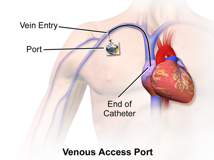
Planting chemotherapy ports
It is the placement of a special stent under the skin and at the top of the chest for easy, pain-free injection of chemotherapy drugs in order to avoid the destruction of the vessel.
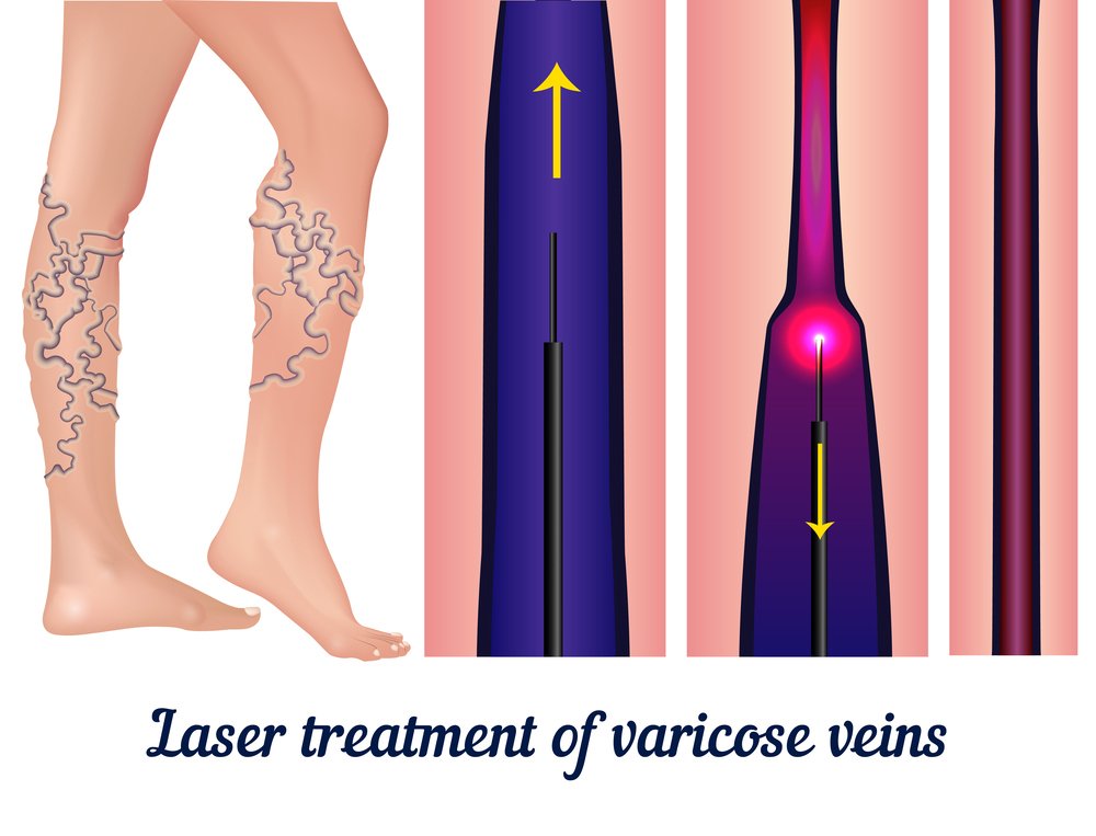
Laser of varicose
Laser of varicose is another non-surgical method for the treatment of varicose. In this method, the vessels path will be obstructed by fiber laser passing through the defective vessels and radiating laser. Then, this area will be removed from the blood supply network and, therefore, varicose will be treated.
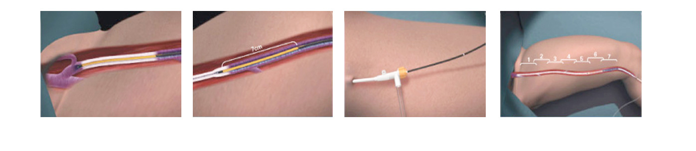
Radiofrequency obliteration of the varicose vein of lower limb
This technique is another non-surgical procedure that became common after laser. It is the ablation of the internal wall of the defective blood vessel by catheter and through radiofrequency waves. In this method, the patient feels less pain than laser.
Endoscopic disc
It is a minimally invasive technique in which endoscope is inserted into a ruptured disk space removing the parts that exert pressure on the spinal cord and releasing the spinal canal.
Disc laser
This method is suitable for mild ruptured disc in which rupture in the disc herniation space will be repaired by placing fiber laser and radiating laser with the help of CT scan.

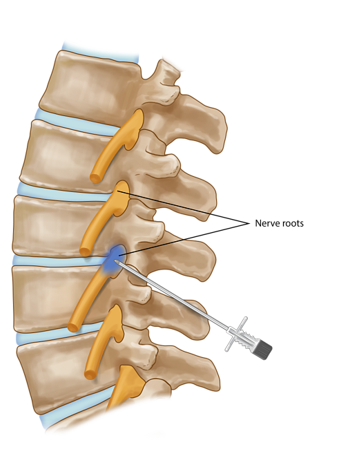
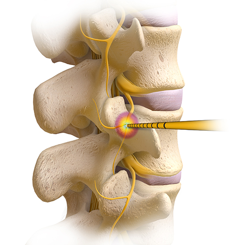
Nerve root injection (PRT)
In this method, with the help of CT scan and using a long thin needle, a combination of some special anti-inflammatory and sedative drugs will be injected to the involved area of the nerve root. This should be performed 3 to 4 sessions every 3 weeks. If no reply is obtained, there is no need for continuing the treatment.
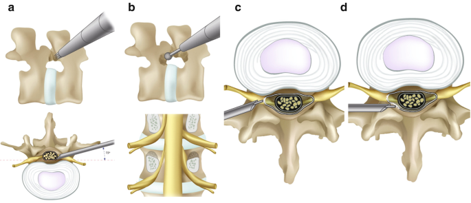
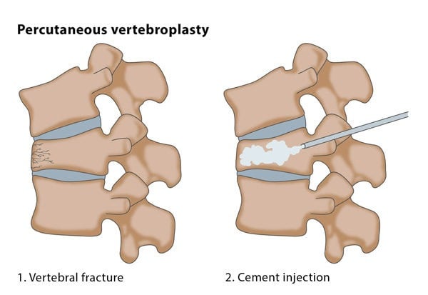
Vertebroplasty
In some cases, osteoporosis or tumors in lumbar vertebrae destroy the vertebral body, causing severe pain. In this method, using CT fluoroscopy, a special cement will be injected into the destroyed vertebra and will strengthen fractured vertebra and the patent’s pain disappears immediately after the surgery.
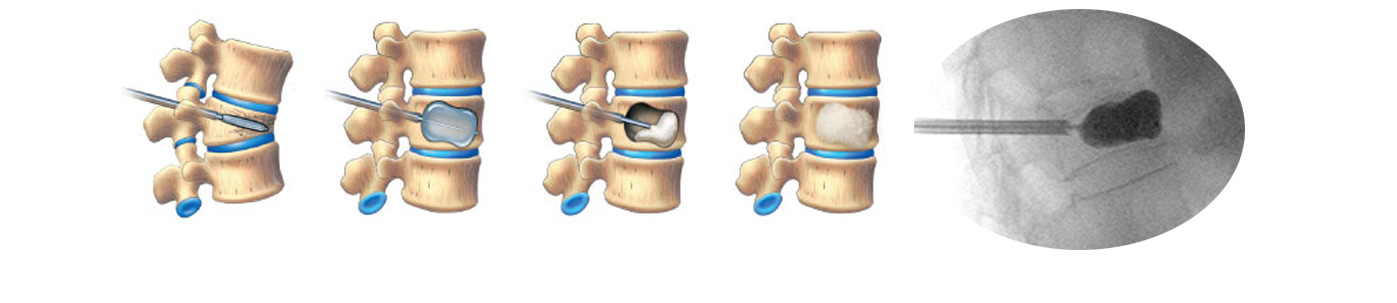
Kyphoplasty
To strengthen the compressed vertebra, the vertebra height will be first adjusted by the balloon and, then, the cement will be injected into the cavity in the vertebra.
Radiofrequency of disc herniation
In this method, disc rupture or disc herniation will be repaired by radio frequency waves tear under the direction of the imaging device.
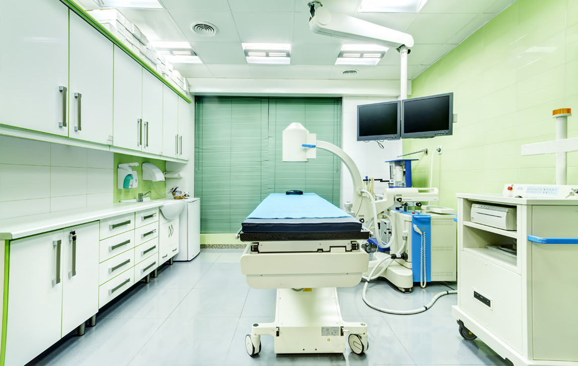
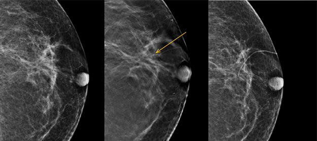
Marking breast lumps
Some breast lumps are not easily available during the surgery. In this case, the radiologist will find the lump using the imaging device and places a very thin spring inside the lump. The other end of this spring will be left out of the breast skin for easily bringing out the lump
Double J stent
Ureteral obstruction by tumor or post-surgery adhesion leads to the accumulation of urine in the kidneys (hydronephrosis) and increased blood creatinine level. To remove the ureteral obstruction, the stent will be inserted into the stenosis using the imaging device, which connects the urinary path with urinary bladder and removes the obstruction.

Microwave ablation of the liver, lung, kidney, and other lesion
The is a new microwave tumor ablation technique. It is more suitable for the ablation of large tumors near the blood vessels and is used for the ablation of tumors of the liver, lungs, etc.
Sclerotherapy of spider varices (telangiectasia)
Thin capillaries scattered irregularly at the surface of lower limbs, which are not aesthetically appropriate, are treated by the injection of sclerosing into the arteries.
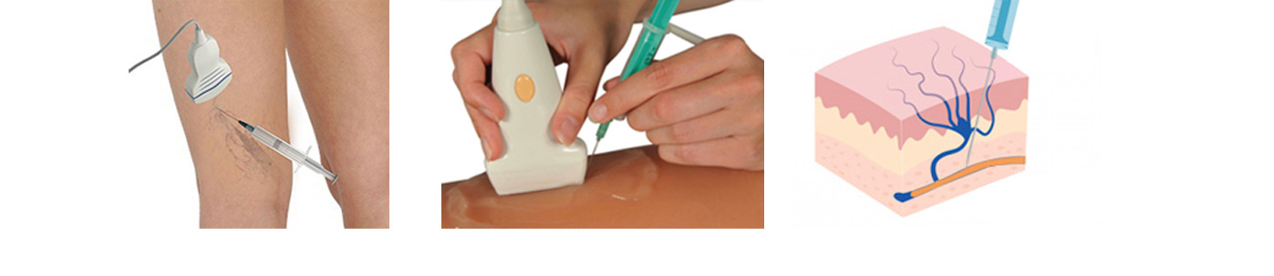
Foam sclerotherapy
Varicose veins with a diameter greater than 1.5 mm are treatable through foam sclerotherapy method. That is, through injection of a special drug into the blood vessels, the defective vessels are blocked, as a result of which treatment will be achieved.
Dilatation of the bile ducts
In some patients, despite stenting and catheterization of biliary duct obstruction, the biliary duct through which bile is discharged into the intestine is still obstructed due to that tumor growth. In these situations, obstruction will be opened by angioplasty balloon and the bile can be easily discharged to the intestine
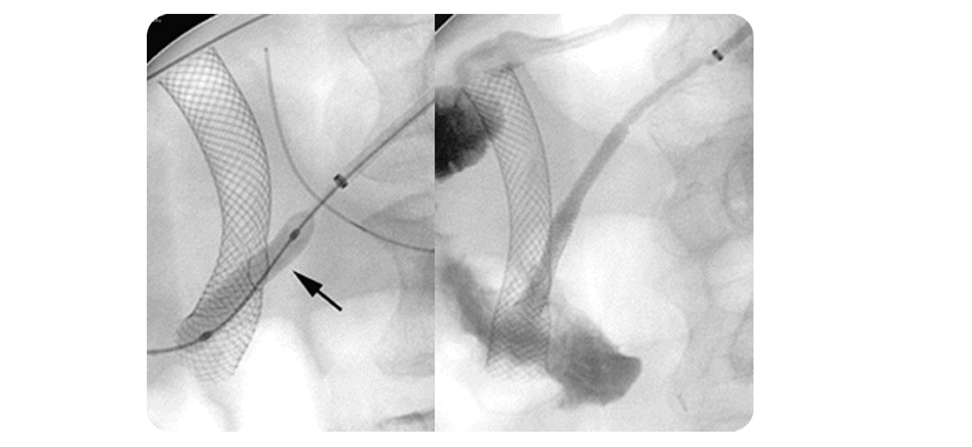
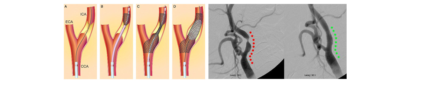
Stenting narrowed carotid arteries, vertebral arteries, etc.
In this procedure, special springs are placed for unblocking vessels. It is performed using an imaging device and through angiography. That is, a special catheter is inserted into the blocked area through the groin right and, then, vessels will be dilated by angioplasty balloon and the special spring (stent) will be inserted into the stenosis.
Direct chemotherapy of retinoblastoma tumor via ophthalmic artery
In order to prevent enucleation owing to this type of tumor, a combination of a few types of chemotherapy drugs will be injected into the artery feeding the eye tumor (ophthalmic artery). This procedure has had very significant results and is performed using an imaging device and through angiography.