mammography
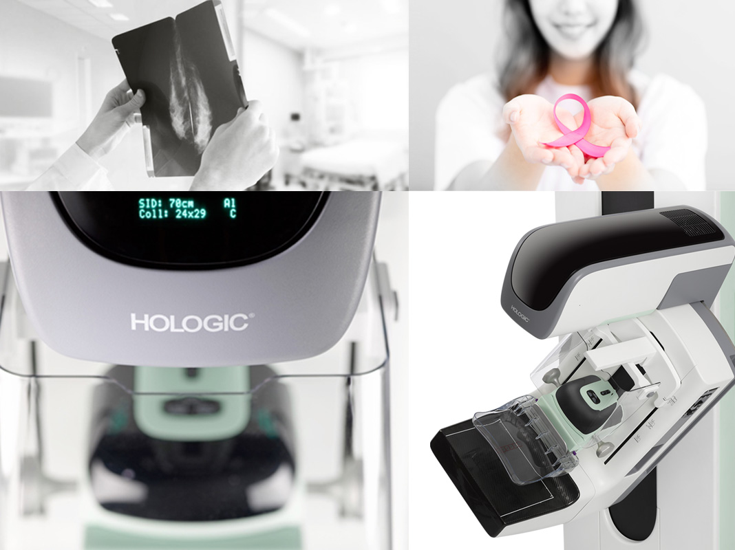
Mammography is one of the most important, effective ways for detecting breasts cancer, especially in the early stage of the disease. One of the most effective ways for fighting this disease in to diagnose it in the early stages.
Mammography is the radiography of the breast soft tissue, mainly used for detecting and diagnosing breast cancer and also for evaluating the palpable lumps and non-palpable lesions of the breast.
Women over forty are recommended to perform mammography once in two years. The women, whose immediate relatives (mother or sister) have a history of breast cancer, are recommended to perform mammography annually from the age of 35. The best time for taking a mammogram is the first week of the period.
It is recommended that:
You should inform he technologist if you have breast implants, so that the appropriate imaging method would be used.
When you go for a mammography, make sure to have your previous mammography images with you because for definitive diagnosis, it is necessary to compare the present mammography images with the previous ones.
Application of mammography
Screening of women over 40 for breast cancer
Evaluation of patients with suspicious breast lumps
Treatment of patients under radiotherapy or mastectomy
Monitoring the contralateral breast in patients who have removed their cancerous breast
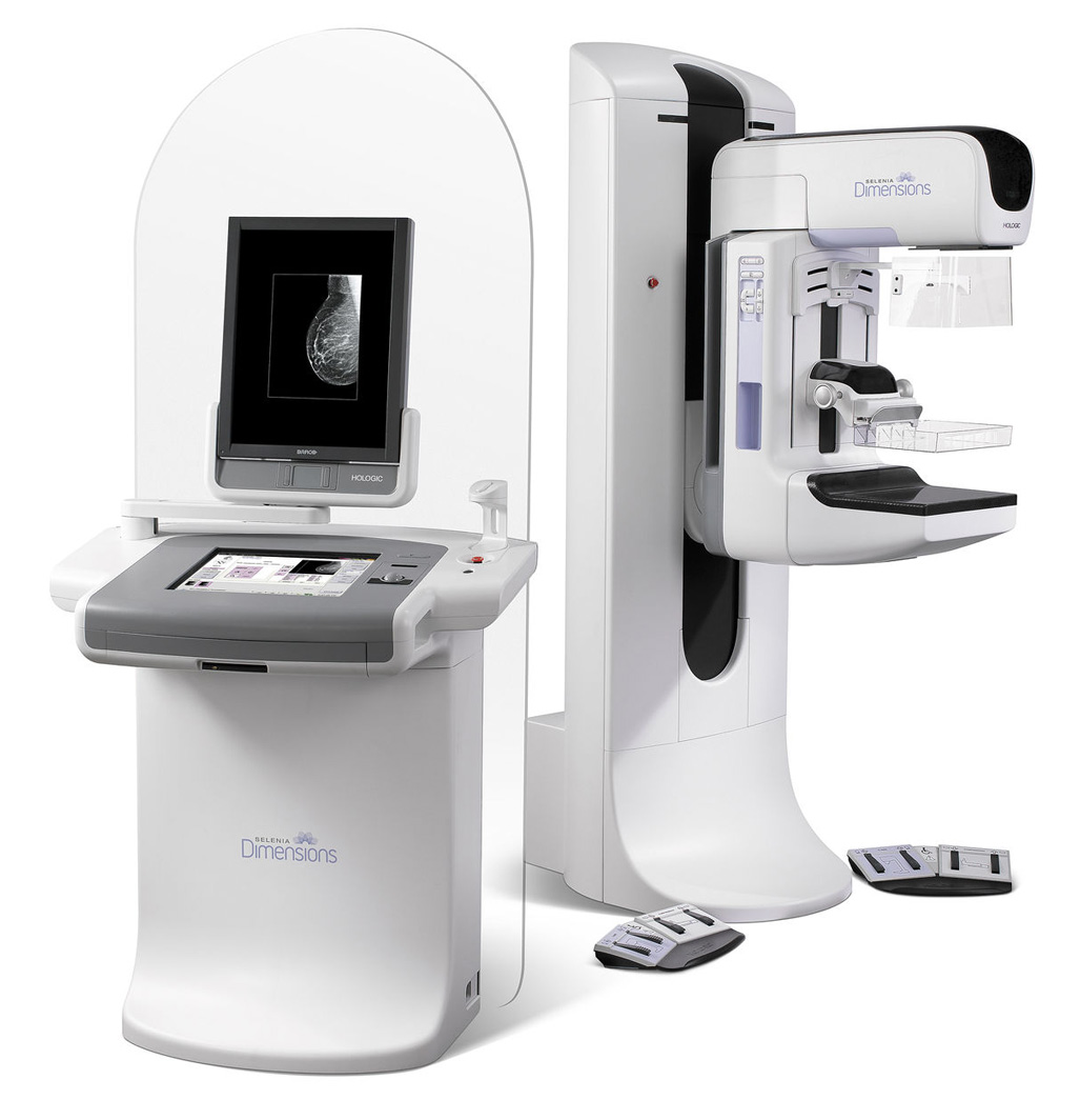
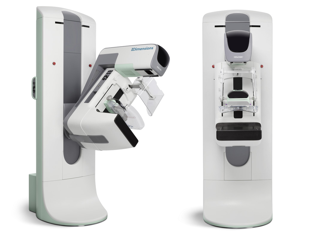
Digital Mammography
How mammography is performed
In mammography, the patient stands in front of a mammography device so that the patients breast could be placed between two plates of the mammography device. These two plates compress the breast to perform imaging. Then, the breast image will be created by radiating X-ray to the breast tissue. Two images of each breast are taken, one after pressing the breast horizontally and the other one after pressing it vertically.
When you go for mammography, do not use antiperspirant spray, lotion, or talcum powder on your armpit or breasts because their traces will be seen as calcium deposits (micro calcification) in the mammography images that interfere with the accurate, precise diagnosis of the disease.